Breaking Barriers: 50-Hour Tuberculosis Test, Migrating Neuron DNA Breaks, and Programmable Virtual Metalenses
Breaking Barriers: 50-Hour Tuberculosis Test, Migrating Neuron DNA Breaks, and Programmable Virtual Metalenses
This week has seen remarkable breakthroughs across the physical, biological, and engineering sciences, demonstrating the power of molecular-level manipulation. From a rapid single-cell diagnostic that slashes tuberculosis resistance testing from two months to two days, to the discovery that migrating brain cells routinely repair double-strand DNA breaks caused by mechanical stress, scientists are redefining biological boundaries. Meanwhile, optical engineers have created a programmable "virtual metalens" that dynamically upconverts infrared light to the visible spectrum, promising cheap, lightweight, and software-adjustable night-vision and thermal imaging.
🔬 Rapid Diagnostics: 50-Hour Phenotypic Drug Susceptibility Testing for Tuberculosis
Tuberculosis (TB) remains one of the world's deadliest infectious diseases, with drug-resistant strains posing a catastrophic challenge to global healthcare systems. Traditionally, determining whether a patient's TB infection is resistant to key antibiotics—known as phenotypic drug susceptibility testing (pDST)—has been a slow and tedious process. Because Mycobacterium tuberculosis is exceptionally slow-growing, clinicians must culture the bacteria in a broth for four to eight weeks before they can observe whether a drug inhibits cell growth. This delay means patients are often placed on broad-spectrum, toxic, or ineffective drug regimens while waiting for results, fueling the spread of multi-drug-resistant TB (MDR-TB).
To solve this critical bottleneck, a collaborative research team including scientists from the Suzhou Institute of Biomedical Engineering and Technology (SIBET) under the Chinese Academy of Sciences, the University of Science and Technology of China (USTC), and the University of Oxford have developed a revolutionary method called Raman-Deuterium Isotope Probing (Raman-DIP). Published in Analytical Chemistry on June 19, 2026, their study demonstrates that this single-cell phenotypic testing method can identify antibiotic resistance and determine minimum inhibitory concentrations (MICs) in just 50 hours—less than 10% of the time required by traditional methods.
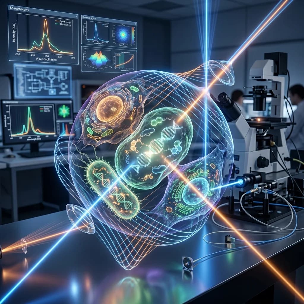
The physics behind Raman-DIP is as elegant as it is precise. The researchers incubate M. tuberculosis cells in a solution containing heavy water ($D_2O$, where hydrogen is replaced with its heavier isotope, deuterium) along with first-line TB antibiotics (rifampicin, isoniazid, streptomycin, and ethambutol). Living, metabolically active bacteria absorb the heavy water and incorporate deuterium into their newly synthesized carbon-hydrogen bonds, forming carbon-deuterium ($C-D$) bonds. Single-cell Raman spectroscopy then detects these $C-D$ bonds, which vibrate at a unique frequency. If a drug successfully disables the bacteria, their metabolism halts, and no $C-D$ signal appears. If they are drug-resistant, they continue to metabolize and generate a strong Raman signature. Because this metabolic measurement is done at the single-cell level, it bypasses the need to wait weeks for cells to replicate, delivering highly accurate, culture-free results in just over two days.
🧠 Cellular Choreography: How Migrating Neurons Survive Routine DNA Breaks
During embryonic development, newborn neurons must travel long distances from their birthplaces deep within the brain to their final destinations in the outer layers of the cerebral cortex. This process, known as neuronal migration, is essential for building the complex, layered architecture of the brain. To get there, these young cells must squeeze through extremely narrow, crowded intercellular spaces. A groundbreaking study published in Nature on June 17, 2026, has revealed a startling consequence of this journey: the physical compression neurons experience during migration actually tears their genomic DNA, causing double-strand breaks (DSBs) that are rapidly and safely repaired once they reach their destination.
The research was led by Zhejing Zhang and Mineko Kengaku at Kyoto University’s Institute for Integrated Cell-Material Science (WPI-iCeMS), in collaboration with scientists from Nagoya University, Tokyo University, and the National University of Singapore. Double-strand DNA breaks are typically considered highly dangerous events; in normal cells, they trigger programmed cell death (apoptosis) or cause chromosomal rearrangements that lead to cancer. However, the Kyoto-led team found that in developing cortical neurons, these breaks are a routine and non-lethal physiological consequence of migration.
To study this phenomenon, the researchers tracked migrating cortical neurons in mouse embryos and designed microfluidic channels with narrow constrictions as tight as 1.5 micrometers to simulate brain tissue in vitro. They observed that as a neuron squeezes its nucleus through these tight bottlenecks, the mechanical stress physically deforms the nuclear envelope, causing chromatin to stretch and snap, leading to double-strand breaks. Fortunately, these neurons have evolved a highly efficient response. As they migrate, they upregulate DNA damage response machinery. Once the cells reach the cortical plate and stop migrating, they rapidly initiate Non-Homologous End Joining (NHEJ)—a quick-repair pathway that stitches the broken DNA ends back together. This discovery not only sheds light on how the brain builds itself but also suggests that incomplete or imperfect DNA repairs during migration could introduce tiny, localized somatic mutations (somatic mosaicism). This genetic diversity among individual neurons may contribute to the unique wiring and complexity of the human brain.
👁️ Nonlinear Optics: Programmable "Virtual Metalenses" for Infrared Imaging
Infrared (IR) imaging is indispensable for a wide range of applications, including military night vision, medical diagnostics, agricultural monitoring, and the collision-avoidance systems of autonomous vehicles. However, standard infrared detectors are expensive, bulky, and often require cryogenic cooling to eliminate thermal noise. Furthermore, traditional optical lenses are static; their focal length and magnification are permanently set during manufacturing. In a significant leap for integrated optics, researchers at Nottingham Trent University (NTU) have developed a "virtual metalens" that can dynamically shape and upconvert infrared light into visible wavelengths, allowing standard, inexpensive visible-light cameras to capture high-resolution multispectral infrared images.
Led by Distinguished Professor Mohsen Rahmani and published in Advanced Photonics Nexus in early July 2026, the study introduces a reconfigurable imaging system that replaces traditional physical lenses with a laser-driven "virtual" lens. Instead of etching fixed nanostructures onto glass—the approach used in conventional metalenses—the team utilized a Spatial Light Modulator (SLM) to project dynamically adjustable phase patterns onto a pump laser beam. This pump beam is then combined with incoming infrared light on a nonlinear optical crystal.
graph LR
    IR[Infrared Light from Object] -->|Focuses onto| NC[Nonlinear Crystal]
    Laser[Pump Laser Beam] -->|Passes through| SLM[Spatial Light Modulator]
    SLM -->|Creates Holographic Phase Pattern| VL[Virtual Metalens Pump Beam]
    VL -->|Combines with IR| NC
    NC -->|Sum-Frequency Generation SFG| Vis[Visible Light Output]
    Vis -->|Captured by| CMOS[Standard Visible CMOS Sensor]
Within the nonlinear crystal, a quantum process called Sum-Frequency Generation (SFG) takes place. The crystal merges the incoming infrared photons with the pump beam's photons, generating new photons whose frequency is the sum of the two inputs. This shifts the light from the invisible infrared spectrum into the visible range. Because the pump beam carries the phase information of the virtual lens programmed into the SLM, the newly created visible light is focused directly onto a standard silicon CMOS sensor (like the one in a smartphone camera). By changing the software code controlling the SLM, researchers can alter the focal length, zoom, and select specific infrared wavelengths almost instantaneously, without any moving parts. This software-defined approach could democratize infrared imaging, making high-end night-vision and thermal sensing lightweight, inexpensive, and highly adaptable.
📊 Comparative Summary of Breakthroughs
| Technology Domain | Traditional Method / Limitation | Breakthrough Innovation | Primary Advantage |
|---|---|---|---|
| TB Diagnostics | Broth-based culture (requires 4–8 weeks for cell growth) | Raman-Deuterium Isotope Probing (Raman-DIP) | Results in 50 hours at single-cell level |
| Neurodevelopment | DNA double-strand breaks considered purely pathological/lethal | Migrating neurons undergo transient, repairable DSBs | Facilitates cortical positioning & somatic diversity |
| Infrared Optics | Expensive, static lenses & cooled semiconductor detectors | Reconfigurable "Virtual Metalens" via SLM upconversion | Real-time programmable focal length; visible CMOS capture |
📌 The Bottom Line
- tuberculosis-raman-dip: The Raman-DIP method cuts tuberculosis drug resistance testing time from weeks to just 50 hours, allowing rapid personalization of lifesaving antibiotic regimens.
- neuron-migration-dna-breaks: Squeezing through narrow pathways during brain development causes physical double-strand breaks in neuronal DNA, which are rapidly repaired to ensure survival and potentially enhance brain cell diversity.
- programmable-virtual-metalens: The virtual metalens dynamically converts infrared light into visible light using a programmable laser, allowing standard visible-light cameras to perform software-controlled thermal and night-vision imaging.
References & Scientific Literature:
- Mao X., et al. "Rapid Phenotypic Drug Susceptibility Testing of Mycobacterium tuberculosis Using Raman-Deuterium Isotope Probing." Analytical Chemistry, June 19, 2026. DOI: 10.1021/acs.analchem.6c01283.
- Zhang Z., et al. "Confined migration induces non-lethal DNA damage in developing neurons." Nature, June 17, 2026. DOI: 10.1038/s41586-026-06830-w.
- Zheng Z., Xu L., & Rahmani M. "Nonlinear virtual lens for programmable and multispectral infrared upconversion imaging." Advanced Photonics Nexus, July 2, 2026. DOI: 10.1117/1.APN.3.4.046002.
Enjoyed this post?
Get our weekly digest delivered free.
Share this post:
Knowelth is reader-supported. We may earn a commission from links in this article at no extra cost to you. Read our disclosure.
Related Posts

Targeted Protein Degrader Rewrites Leukemia Drug Sensitivity, Oxytocin Selectively Modulates Low Trust, and Siberian Permafrost Methane Surge Doubles
Targeted Protein Degrader Rewrites Leukemia Drug Sensitivity, Oxytocin Selectively Modulates Low Trust, and Siberian Permafrost Methane Surge Doubles Scientific discovery rarely moves in a single dire…
Global Research Breakthroughs: HCMI Patient Cancer Organoids, Ultra-Durable Nanochannel Fuel Cells, and 567-Million-Year-Old Deep-Ocean Fossils
Global Research Breakthroughs: HCMI Patient Cancer Organoids, Ultra-Durable Nanochannel Fuel Cells, and 567-Million-Year-Old Deep-Ocean Fossils Across biology, materials physics, and evolutionary pale…
Tumour Organoid Vulnerability Map, 567M-Year-Old Ediacaran Fossils, and NIH's TrialGPT AI Breakthrough
Tumour Organoid Vulnerability Map, 567M-Year-Old Ediacaran Fossils, and NIH's TrialGPT AI Breakthrough Three landmark studies published in the first week of August 2026 span the full breadth of modern…